

Removal of fibromas and Malherbe epitheliomas
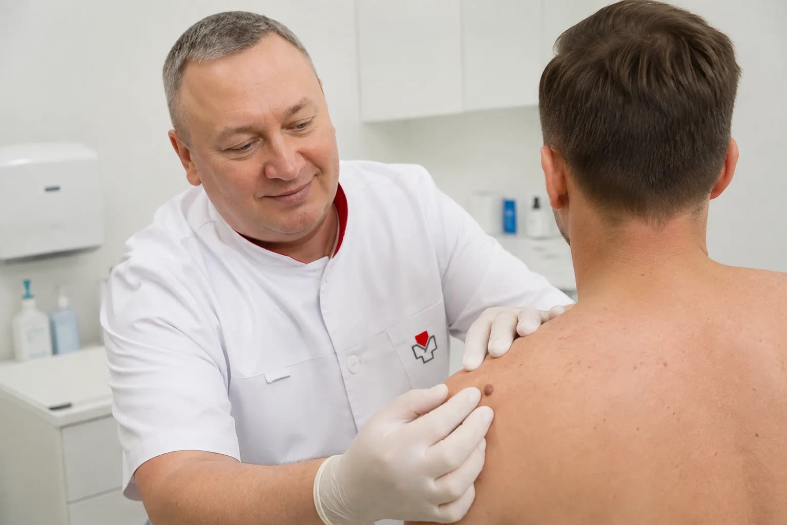
Fibromas and Malherbe epitheliomas are benign lesions that may develop in the skin or subcutaneous tissues of the trunk and limbs. During the examination, the surgeon assesses their size, location, and clinical characteristics and determines whether surgical removal is indicated and which approach is appropriate.

Specialists
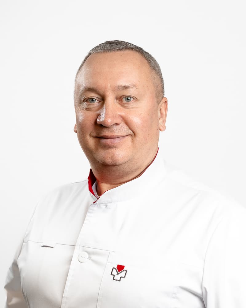
Bosyy Oleh Anatoliyovych
Ultrasound doctor, Surgeon
Fibromas and Malherbe epitheliomas: what are they and why do they develop?
Fibromas and Malherbe epitheliomas are benign lesions with different origins. They are often noticed as a “lump” or an area of thickening that has appeared under the skin and does not disappear. However, the exact type of a lesion cannot be determined by appearance alone, as cysts, lipomas, and other changes in the skin and subcutaneous tissues may present in a similar way.
A fibroma is a lesion associated with connective tissue. Depending on its type, it may be soft or firm and may develop on the surface of the skin or deeper within the tissues. Some fibromas remain unchanged for years, while others gradually increase in size or are repeatedly irritated by clothing or movement.
A Malherbe epithelioma, or pilomatricoma, develops from cells of the hair follicle. It typically presents as a well-defined, firm nodule. Due to calcification within the lesion, it may feel hard to the touch, while the skin over it can sometimes develop a bluish, reddish, or uneven appearance. The lesion usually grows slowly and does not disappear on its own.
The exact cause of pilomatricoma is often unknown. Its development is associated with changes in the cells of the hair follicle. There is also no single universal cause of fibromas, as this depends on the specific type of lesion and the tissue from which it develops.
An important point is that not every lump needs to be removed immediately, but any new or changing lesion should be properly assessed. This is particularly important if it is increasing in size, becoming painful, changing colour, being repeatedly traumatised, or causing discomfort because of its location.
During a surgeon consultation, the doctor assesses the size, consistency, mobility, and location of the lesion. Additional diagnostic tests may be recommended when necessary. After surgical removal, the tissue may be sent for histological examination, which allows the structure and nature of the lesion to be determined more precisely.
FAQ
How does the removal procedure work, and what should you expect during healing and aftercare?
Before removal, the surgeon examines the lesion and determines the extent of the procedure. The procedure is usually performed under local anaesthesia. Once the area is numb, the surgeon makes an incision, removes the lesion, and, depending on the size and depth of the wound, places sutures. When indicated, the removed tissue may be sent for histological examination.
After the procedure, the wound is covered with a sterile dressing. The doctor provides individual recommendations regarding dressing changes, contact with water, physical activity, and any other necessary restrictions. Sutures should not be removed independently, and the recommended aftercare routine should not be changed without consulting the doctor.
During the first few days, mild pain, swelling, or slight redness around the treated area may occur. Healing depends on the location and size of the lesion, the depth of the wound, and individual factors. If the pain increases, redness spreads, discharge appears from the wound, or any other unusual changes occur, contact your doctor before the scheduled follow-up appointment. If sutures were placed, they are removed at a time determined by the surgeon, depending on the location of the wound and the healing process.
Booking a consultation and procedure
If the lesion requires assessment or you are already considering its removal, book a consultation with a surgeon. During the appointment, the surgeon will evaluate the lesion, determine the appropriate scope of the procedure, and explain the next steps.
To schedule a consultation or procedure, call +380 97 312 92 91. Doctor Sam is located at Respublika Park Shopping Mall, 1 Kiltseva Road, Kyiv, near Teremky Metro Station.


