

Transabdominal pelvic ultrasound
Ultrasound examination helps assess the condition of the pelvic organs, identify possible abnormalities and investigate the causes of certain symptoms. It may be recommended for lower abdominal pain, menstrual irregularities, unusual vaginal bleeding, routine examinations and monitoring previously identified changes.
What is a transabdominal pelvic ultrasound?
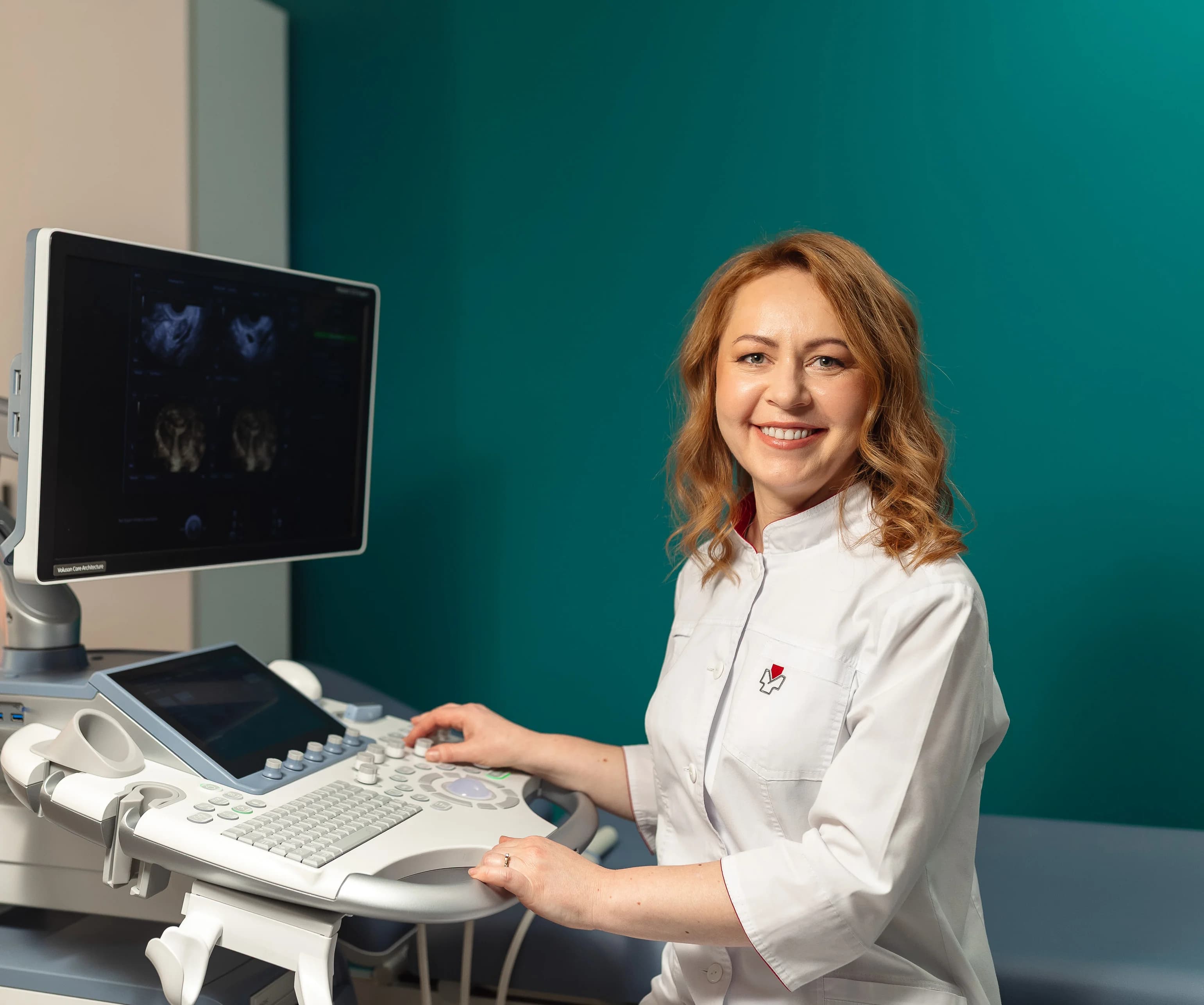
A transabdominal pelvic ultrasound is an imaging examination in which a healthcare professional examines the pelvic organs through the lower abdominal wall using an external probe. The probe is not inserted into the vagina, making this approach a suitable option for women who prefer or require an external examination.
During the scan, the doctor assesses the position, size and structure of the uterus and ovaries, as well as other organs within the examination area. The scan can help identify changes such as uterine fibroids and ovarian cysts, and investigate possible causes of lower abdominal pain, unusual vaginal bleeding or menstrual irregularities.
The examination uses ultrasound waves and does not involve ionising radiation. If a more detailed assessment is needed, your doctor may recommend additional imaging, including a transvaginal ultrasound.

Specialists

Halenko Rymma Oleksandrivna
Ultrasound doctor, Obstetrician-gynecologist

Yarosh Oksana Ivanivna
Obstetrician-gynecologist, Ultrasound doctor

Oliinyk Anna Oleksandrivna
Obstetrician-gynecologist, Ultrasound doctor

Nosko Alla Oleksandrivna
Ultrasound doctor

Zahrebelna Liudmyla Hryhorivna
Obstetrician-gynecologist, Ultrasound doctor

Bosyy Oleh Anatoliyovych
Surgeon, Ultrasound doctor

Lytvyn Yana Yuriivna
Ultrasound doctor, Pediatric gynecologist, Obstetrician-gynecologist
How to prepare for a pelvic ultrasound
For a transabdominal pelvic ultrasound, your bladder should be full. This helps produce clearer images of the pelvic organs. You are generally advised to drink approximately 500–700 ml of still water about an hour before your appointment and avoid emptying your bladder until the examination is complete.
You usually do not need to change your diet or fast before a pelvic ultrasound performed on its own. Do not change your regular medication schedule unless instructed by your healthcare professional. If your appointment includes other types of ultrasound, the preparation requirements may differ.
Additional examinations and consultations
- Obstetrics and gynaecology
- Colposcopy
What can be assessed during a pelvic ultrasound?
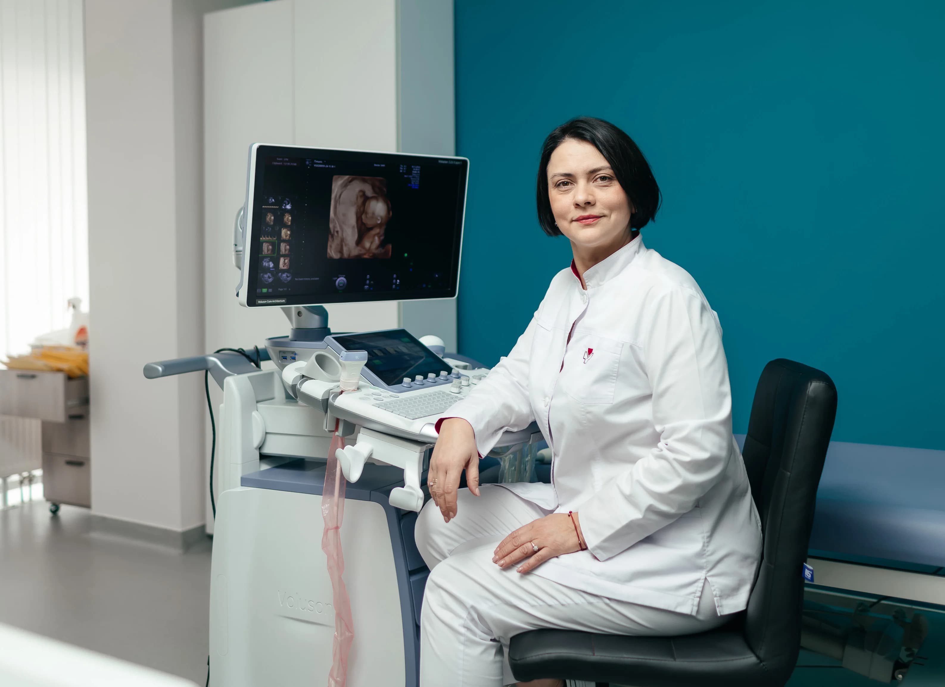
During the examination, the doctor assesses the position, size and structure of the uterus, the endometrium (the inner lining of the uterus) and the ovaries. Particular attention is paid to any masses, tissue changes or other findings that may require further investigation.
A pelvic ultrasound can identify uterine fibroids, ovarian cysts and other structural abnormalities. The results help the doctor determine whether additional examinations are needed to clarify the diagnosis.
Frequently asked questions
Can I have a pelvic ultrasound during my period?
Which day of my menstrual cycle is best for a pelvic ultrasound?
Can I have a transabdominal ultrasound if I have never been sexually active?
What is the difference between transabdominal and transvaginal ultrasound?
Is a pelvic ultrasound painful?
Is ultrasound imaging safe?


